Ocular Surface Disease

Ocular surface disease (OSD) encompasses a range of disorders affecting the surface of the eye, which consists of the cornea and conjunctiva. These conditions can significantly impact an individual’s quality of life, causing eye irritation, pain, tearing, blurry vision, and even corneal abrasions and infections. Understanding the symptoms, diagnostic methods, and treatment options for OSD is crucial for patients and healthcare providers alike.

The causes of OSD can be divided into several broad groups, some of which can overlap: (1) insufficient or unstable tear film (i.e., “dry eye”), (2) poor epithelial healing related to stem cell dysfunction, (3) poor epithelial healing related to a nerve disorder, and (4) acute or chronic tissue inflammation. Patients can manifest one or more of these groups of conditions and findings, which is what makes treatment of OSD so challenging: many patients require individualized treatment of each factor contributing to their disease.

We perform the following treatments:

  • Amniotic membrane placement
  • iLux and BlephEx for dry eyes and blepharitis
  • Reconstructive surgery for ocular cicatricial pemphigoid, Stevens Johnson Syndrome, toxic epidermal necrolysis, burns, and more

Symptoms of Ocular Surface Disease

Ocular surface disease can manifest through a variety of symptoms, often depending on the specific condition involved. Common symptoms include:

  • Dryness: A sensation of dry eye is a hallmark of ocular surface disease, often resulting from inadequate tear production or poor tear quality.
  • Redness: Inflammation and irritation can cause the eyes to appear red and bloodshot. The redness is caused by dilation of conjunctival and sub-conjunctival blood vessels (capillaries) in response to irritation and inflammation.
  • Blurred vision: Fluctuations in vision or persistent blurriness can occur, often as a result of tear film instability.
  • Discomfort or pain: Patients may experience a gritty or scratchy sensation as if something is in their eye (“sand in the eye” sensation).
  • Light sensitivity: Increased sensitivity to light, known as photophobia, is very common because the sensory nerves of the ocular surface can become hypersensitive in the context of chronic irritation.
  • Tearing: Ironically, excessive tearing can be a response to dry eye conditions, as the eye attempts to compensate for the dryness.
  • Recurrent corneal erosions: The superficial part of the cornea (epithelium) can abrade (scratched off) if the surface of the eye is overly dry, especially if corneal sensation has been damaged.

Diagnosis of Ocular Surface Disease

Diagnosing OSD requires a comprehensive eye examination, often involving several tests:

  • Patient history: Understanding the patient’s symptoms, general health, medications, and environmental exposures is crucial.
  • Visual acuity test: This assesses how well the patient can see at various distances, with and/or without glasses.
  • Slit-lamp examination: A microscope examines the structures of the eye in detail, checking for signs of disease on the ocular surface, particularly inflammation and fluid accumulation (“chemosis”).
  • Tear film evaluation: Tests like the Schirmer test measure tear production, while tear film break-up time (TBUT) assesses tear film stability.
  • Corneal staining: Special dyes like fluorescein can highlight surface damage to the cornea and conjunctiva.
  • Meibomian gland evaluation: Dysfunction of these glands can contribute to dry eye and is assessed through gland expression and imaging. The meibomian glands produce specialized oils (“mebum”) that coat the surface of the tear film to lubricate the blink and reduce evaporation. Meibomian gland disease (MGD) is usually caused by chronic inflammation and/or low-grade infection, and can lead to death of glandular cells, resulting in long-term irreversible damage.

Treatment Options for Ocular Surface Disease

Treatment for OSD varies depending on the underlying cause and severity of the condition. Treatments are generally divided into non-surgical and surgical. At Kahana Oculoplastic and Orbital Surgery, we have expertise in all aspects of OSD therapy, including the most advanced surgical interventions. Contact us if you have any questions.

  • Artificial tears: Over-the-counter lubricating eye drops and gels can provide temporary relief for dry eyes. The thicker gels and ointments provide longer-lasting lubrication and protection but can also cause blurry vision.
  • Prescription medications: Anti-inflammatory drugs, such as prednisolone (steroid), cyclosporine or lifitegrast eye drops, can reduce inflammation and improve tear production.
  • Punctal plugs: These small devices, inserted into the tear ducts, can help retain moisture on the eye’s surface by slowing tear drainage.
  • Eyelid hygiene: Regular cleaning of the eyelid margins can help manage conditions like blepharitis and MGD, which often coexist with OSD.
  • Omega-3 fatty acid supplementation: Dietary changes or supplements can sometimes improve tear quality because production of mebum (tear film oils) requires mono-unsaturated fatty acids.
  • Environmental modifications: Adjusting factors like humidity/dry air, wind exposure, and screen time can alleviate symptoms.
  • Specialty contact lenses: In severe cases, lenses like scleral contacts can protect the cornea and trap moisture.
  • Surgery, or ocular surface reconstruction: In more severe or recalcitrant cases, surgical intervention may be necessary to correct structural or anatomic issues contributing to OSD.

Surgical Interventions

At Kahana Oculoplastic and Orbital Surgery, we have several surgical tools to improve the signs and symptoms of OSD, often getting at the heart of what’s causing the OSD.

  • Correction of eyelid malposition: Eyelid conditions such as ectropion (outward turning) or entropion (inward turning) lead to poor protection of the cornea and conjunctiva, poor distribution of tears, lashes rubbing against the cornea, and keratinization of the conjunctiva on the inside of the eyelid (palpebral conjunctiva). Think of the eyelids as being the windshield wipers, and the eye surface as the windshield. If the wipers work poorly, they leave streaks on the windshield that interfere with the view. Likewise with the cornea – if the eyelid blink is poor and the eyelids are poorly fitting, the cornea develops irregularities that interfere with vision. Correcting the eyelid position can improve eye protection, reduce pain and tearing, and visual function. Beyond standard surgical treatments, Dr. Kahana has developed novel techniques that maximize the eyelid’s protective functions.
  • Excise redundant conjunctiva: Conjunctivochalasis is a condition in which the skin of the eye becomes thickened, diseased, and starts rolling over itself with every blink. Each blink should be a smooth movement of the eyelid over the ocular surface. But with conjunctivochalasis, the blink causes the conjunctiva to rub abnormally against the rest of the eye, causing chronic irritation. Identifying and excising the abnormal conjunctiva and reconstructing the eye tissues can greatly improve eye symptoms.
  • Amniotic membrane patching: Ultra-thick amniotic membrane can be sutured onto the surface of the eye to promote regenerative healing of the cornea and conjunctiva. Often the eye needs to be sutured shut for 3-4 weeks while the tissues heal, so this treatment is performed one eye at a time.
  • Lacrimal reconstructive surgery: Sometimes the tear drainage system is blocked or damaged, leading to excessive tearing. In some cases, this contributes to chronic inflammation and even infection of the eye surface, manifesting as mucoid discharge and eye redness. Reconstructive surgery can restore the function of the lacrimal drainage system to improve tear flow and ocular surface health.
  • Corneal neurotization surgery: At Kahana Oculoplastic and Orbital Surgery, we are pioneers in the reinnervation of the cornea using nerve grafting. This is a nerve microsurgery performed under a neurosurgical microscope. We have the most experience with this procedure in the Midwest, but success rates exceed 80% in achieving stabilization of the cornea. Please see section on Neurotrophic Keratitis.

Advanced Therapies

As research progresses, new therapies are emerging:

  • Autologous serum eye drops: Made from a patient’s own blood, these drops contain growth factors that can heal the ocular surface.
  • iLux therapy: This tool is used to treat meibomian gland dysfunction by heating the oil trapped in the glands to an optimal temperature, followed by expressing the oils to clear the glandular apparatus and improve glandular health and function.
  • Regenerative therapy: Amniotic membrane patching uses a non-cellular matrix containing biological factors that can promote natural tissue healing. Brand names include ProKera, which can be placed in the clinic and removed after a few days, or AmnioGuard, which is a surgical graft sutured into position over the ocular surface and stays in place for several weeks.

Ocular surface disease represents a complex set of conditions that require a nuanced approach to diagnosis and treatment. Most patients manifest multiple symptoms that result from multiple causes. In order to achieve a good result with reduction of symptoms, every possible cause of OSD needs to be individually addressed, sometimes sequentially, sometimes simultaneously. At Kahana Oculoplastic and Orbital Surgery, we have a unique combination of skills that covers both the medical and surgical aspects of treatment. We are pioneers in the use of regenerative and reconstructive surgeries to improve eye health. We are proud to be the destination of last resort for patients with OSD refractory to standard therapies.

Corneal Neurotization for Neurotrophic Cornea

An intra-operative photo of nerve fascicles recruited to re-innervate the damaged cornea

The cornea – the clear part in the front of the eye – is a unique biological tissue in that it has no blood vessels and is optically clear. The clarity of the cornea is a fundamental aspect of vision; without this clarity, good vision is impossible.

While the cornea lacks blood vessels and mostly obtains its oxygen supply directly from the air, it is very rich in innervation. It is among the most highly innervated tissues in the body, and even minor injuries to the cornea can be terribly painful (e.g. corneal scratch). Fortunately, the cornea is also the fastest healing tissue in the body. The ability of the cornea to heal requires special biological signals from the nerves. This signals – neurotrophic molecules – stimulate the cells of the cornea to divide and reform the damaged tissue.

A graphic representation of Corneal Neurotization

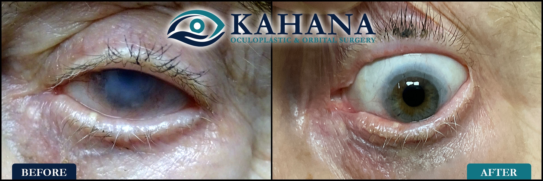
Neurotrophic keratitis is a disorder in which the cornea has lost its nerves. There are many causes to neurotrophic keratitis, including chronic trauma from dry eyes, chronic inflammation, herpes infection, and even congenital absence of the nerve.

A major medical advance of the past decade has been the development of techniques to reinnervate the cornea. These include both medical (eye drops) and surgical (corneal neurotization). The eye drops (cenegermin-bkbj, known as Oxervate) are most useful when the denervation is distal and fairly recent, caused by chronic dry eyes. Otherwise, surgery is required.

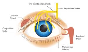
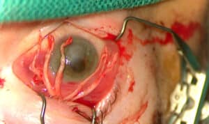
Figures: An intra-operative photo of nerve fascicles recruited to re-innervate the damaged cornea & a graphic representation of Corneal Neurotization

Dry Eyes and Blepharitis

The eye tearing system lubricates and hydrates the eye. The term “dry eye disease” can mean one of two issues. First, aqueous deficient dry eye disease refers to dry eye caused by decreased tear production. Second, evaporative dry eye disease refers to dry eye caused by meibomian gland dysfunction. The signs and symptoms of both affect the entire ocular surface including the tear film, cornea, conjunctiva and eyelids.

Understanding the relationship between dry eyes, ocular surface inflammation, and blepharitis is crucial to treating and maintaining ocular surface health and overall comfort.

Blepharitis refers to inflammation along the base of the lashes and at the meibomian glands along the lid margin. Blepharitis is very common, and people are often asymptomatic for a very long time while the condition progresses. Blepharitis is not a curable condition, however, treatments can alleviate symptoms and prevent the progression of the condition.

Don’t Hesitate to Get in Touch

Don’t hesitate to get in touch if you have any ocular surface disease questions, concerns or issues.