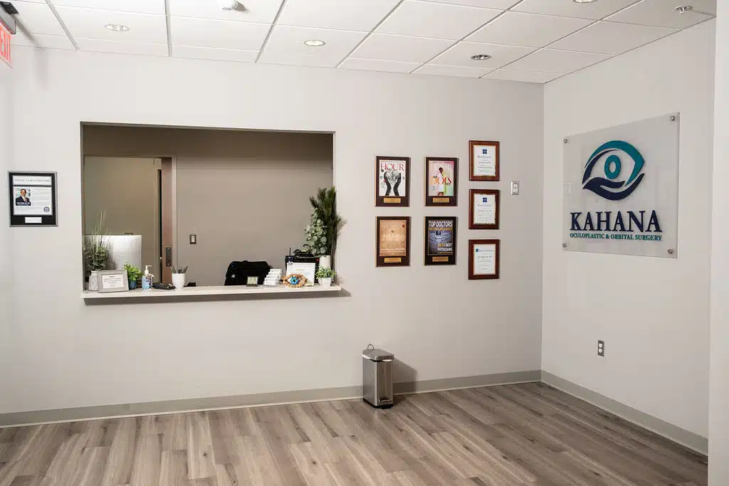
Pediatric Oculoplastic Conditions & Services

Children are not “little adults” and require a unique approach with specialized skills and tools.
We have extensive experience with pediatric oculoplastic disorders, a result of his deep interest in the topic and years of academic research on the developmental biology of the eye and associated structures.
Dr. Kahana has authored numerous manuscripts on the topic of pediatric oculoplastic surgery, taught courses, developed novel techniques, and advanced the science of pediatric oculoplastic surgery.
At birth, vision is not fully developed, and a newborn’s visual acuity is relatively poor. Children’s ability to see improves dramatically over the first 7 years of life, and particularly in the first 2-3 years – the “critical phase of visual development.”
Visual development is driven by brain development in the visual cortex and vision processing centers in the brain. Development of those parts of the brain required for good vision occurs with visual input from the child’s eyes. In Nobel Prize-winning work, Professors David Hubel and Torsten Wiesel of the Johns Hopkins University showed that brain development required visual input from the eyes (view video). If visual input was impaired, then the brain would not develop the ability to see well.
Importantly, there is a critical period of brain development in which the brain can develop the ability to see well. If the visual axis is covered during this time, the brain will never develop the ability to see well – even after the obstruction is removed. Hence, it is very important – even critical – to have a good assessment of visual function by a fellowship-trained pediatric ophthalmologist and to take this issue into account when deciding on when to perform eyelid surgery.
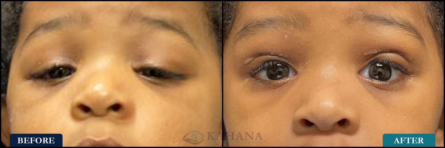
Blepharophimosis syndrome is a rare congenital disorder characterized by a unique set of eyelid malformations.
The primary features include blepharophimosis, a marked reduction in the horizontal width of the eyelids; ptosis, a drooping of the upper eyelids; epicanthus inversus, where the skin fold of the upper eyelid covers the inner corner of the eyes; and telecanthus, an increased distance between the inner corners of the eyes. This constellation of eyelid abnormalities can significantly impact vision and facial appearance.
Children with blepharophimosis syndrome often exhibit a distinctive facial expression and may experience visual impairments due to the ptosis and narrowed palpebral fissures. In some cases, this syndrome is associated with other systemic anomalies or developmental delays. The condition is usually inherited in an autosomal dominant pattern and may require multiple surgical interventions to correct the eyelid abnormalities and improve visual function. Early and comprehensive ophthalmological evaluation is essential for managing the visual aspects and guiding surgical planning to optimize both functional and aesthetic outcomes.

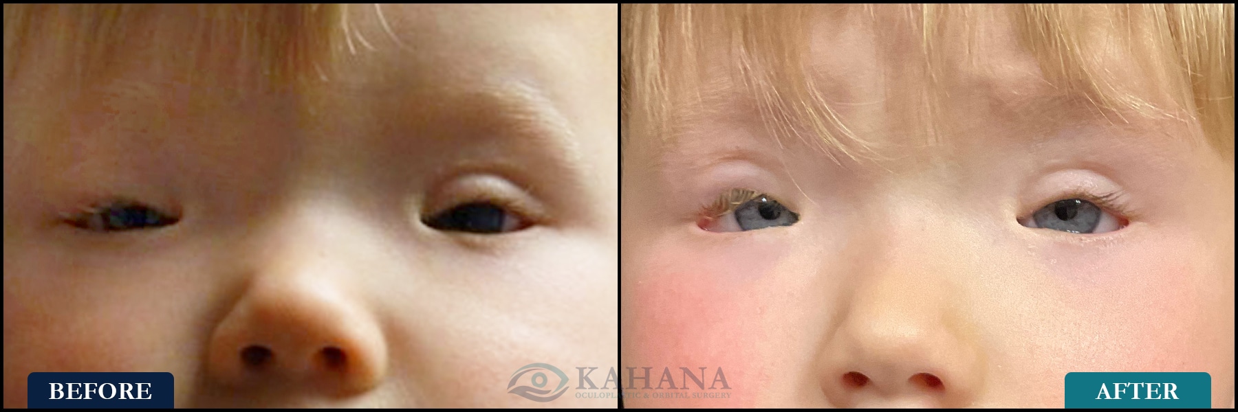

The upper eyelid needs to open sufficiently wide in order to clear the pupil. Weakness of the muscle opening the eyelid (levator muscle) can cause a droopy upper eyelid, known as ptosis.
This can affect both eyes (bilateral) or just one eye (bilateral). When the child needs to raise their chin in order to see from under the eyelid, that is called “ocular torticollis,” which can harm the neck and back muscles and interfere with gross motor development. When the child has unilateral ptosis, the covered eye may be ignored by the brain, resulting in neurologic vision loss, or “amblyopia.”
Correcting ptosis can be done at any age, but the only reason to perform surgery in a very young child is to avoid vision loss or developmental delay. Otherwise, the surgery can wait until the child is at school age.
Eyelid epiblepharon is a condition characterized by an abnormal horizontal fold of skin near the eyelashes, causing the lashes to curve inwards towards the eye.
This condition is most commonly seen in children and is often congenital, particularly prevalent in Asian populations. Unlike entropion, where the entire eyelid margin turns inward, in epiblepharon, the eyelid margin itself remains in a normal position, but the extra fold of skin pushes the lashes into a misdirected path. This can lead to symptoms similar to those of entropion, such as eye irritation, tearing, and redness, as the misaligned lashes rub against the cornea and conjunctiva. However, epiblepharon typically does not carry the same risk of corneal damage as entropion.
In many cases, children outgrow the condition as their facial features develop, though surgical intervention may be required if the symptoms are severe or persist.
The distinction between epiblepharon and entropion is important for proper diagnosis and treatment, since the corrective surgical techniques are different and the latter often requires more immediate and aggressive management.
The tear drainage system includes several distinct anatomic structures that come together (like plumbing) to drain tears from the surface of our eyes. Some infants are born with either an incompletely developed or maldeveloped tear drainage system.
As the child grows, the accumulation of tears and debris within the abnormal drainage system can lead to an infection: dacryocystitis. This manifests as chronic mucoid discharge and crusting along the lashes, as well as tears flowing out of the eye even when the child is not crying.
In addition to a developmental problem in the tear drainage system, some children suffer from malposition of the eyelids or eyelashes, which irritates the eye and causes excess production of tears.
The first step to successful treatment of tearing in children is a complete evaluation and an accurate determination of the cause of the tearing. Once a cause is identified, treatment can range from a particular type of lacrimal sac massage, to surgical reconstruction of the drainage system, to repair of the eyelid malposition. We are passionate about caring for the pediatric population, so if your child has an eyelid or tearing problem, please call us for an appointment. Looking for different types of surgery? Check out our Thyroid Eye Disease and Cosmetic services offerings.
We have extensive experience with pediatric oculoplastic disorders, a result of his deep interest in the topic and years of academic research on the developmental biology of the eye and associated structures.